Her Hair Was Thinning at 37. It Wasn't Genetic
Written and edited by Sarah Bonza, MD, MPH, FAAFP, MSCP, DipABLM, NBC-HWC
What a real hair loss workup looks like for women in perimenopause and midlife.
Short answer for the woman who is scrolling fast: If your hair is thinning in your late 30s or 40s, "it's genetic" and "it's just stress" are diagnoses of exclusion — not starting points. Before either is accepted, six things should be measured: ferritin (with CRP and a full iron panel), thyroid function including antibodies, vitamin D, B12, zinc with copper, and androgens. A normal ferritin is frequently not an optimal ferritin for a hair follicle. That single distinction explains a large share of the women I see who were told nothing was wrong.
Why hair loss lands as an emergency
She was 37. She brought a plastic bag of hair to her appointment — collected from the shower drain over four days, because she was afraid I wouldn't believe her. Her previous visit had lasted eleven minutes and ended with "everyone sheds, it's probably stress, try to relax."
That is not a diagnosis. That is a scheduling constraint wearing a lab coat.
I want to name something clinicians often skip. Hair loss is not cosmetic. For women in midlife, hair is bound up with identity, professional presence, sexual confidence, and the private terror of visibly aging faster than you expected. Women describe hair loss with language they reserve for grief. When a woman is told to relax about it, she hears that her distress is the problem — rather than the shedding.
And "stress" is a non-answer for a mechanical reason: stress-triggered telogen effluvium has a shape. It follows an identifiable insult by roughly two to three months, it sheds diffusely, and it resolves within six to nine months. Shedding that has continued for over a year, or that is accompanied by widening part width, is not a stress story. It is an unfinished workup.
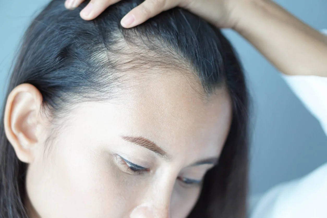
For women in midlife, hair is bound up with identity, professional presence, sexual confidence, and the private terror of visibly aging faster than you expected.
Ferritin: a normal ferritin is not an optimal ferritin
This is the single highest-yield concept in this article.
Most U.S. labs flag ferritin as abnormal below roughly 10–15 ng/mL, because that threshold was built to detect anemia. Hair follicles are among the most metabolically demanding tissues in the body — the matrix keratinocyte is one of the fastest-dividing cell populations we have — and they appear to fail at iron availability well above the anemia threshold.
Rushton's work proposed a target above 70 µg/L for women with increased shedding, explicitly paired with a normal inflammatory marker — a serum ferritin of 70 µg/L alongside a normal erythrocyte sedimentation rate [1]. Kantor and colleagues subsequently confirmed lower ferritin across female hair loss populations [2]. A 2022 systematic review and meta-analysis of 36 studies and just over 10,000 participants found that women with nonscarring alopecia had meaningfully lower ferritin than women without it, and concluded that women with hair loss may benefit from higher ferritin levels [3].
I will be honest about the disagreement, because you deserve it. Olsen and colleagues at Duke studied 381 women and did not find iron deficiency more common in female pattern hair loss or chronic telogen effluvium than in controls [4]. That paper drew a pointed methodological rebuttal from Rushton, Bergfeld, Gilkes, and Van Neste regarding non-standardized sampling and the absence of an inflammatory marker in all subjects [5]. The threshold hypothesis — that iron below a certain level triggers loss only in predisposed follicles — remains unproven by randomized trial [6].
So here is my actual position, stated plainly: I treat ferritin as a modifiable variable, not as a diagnosis. In a woman with active shedding I aim for 70–100 ng/mL, and I tell her honestly that this target is clinical judgment informed by imperfect evidence, not settled fact. The downside of correcting iron in a genuinely low-normal woman is small. The downside of ignoring it for three years is not.
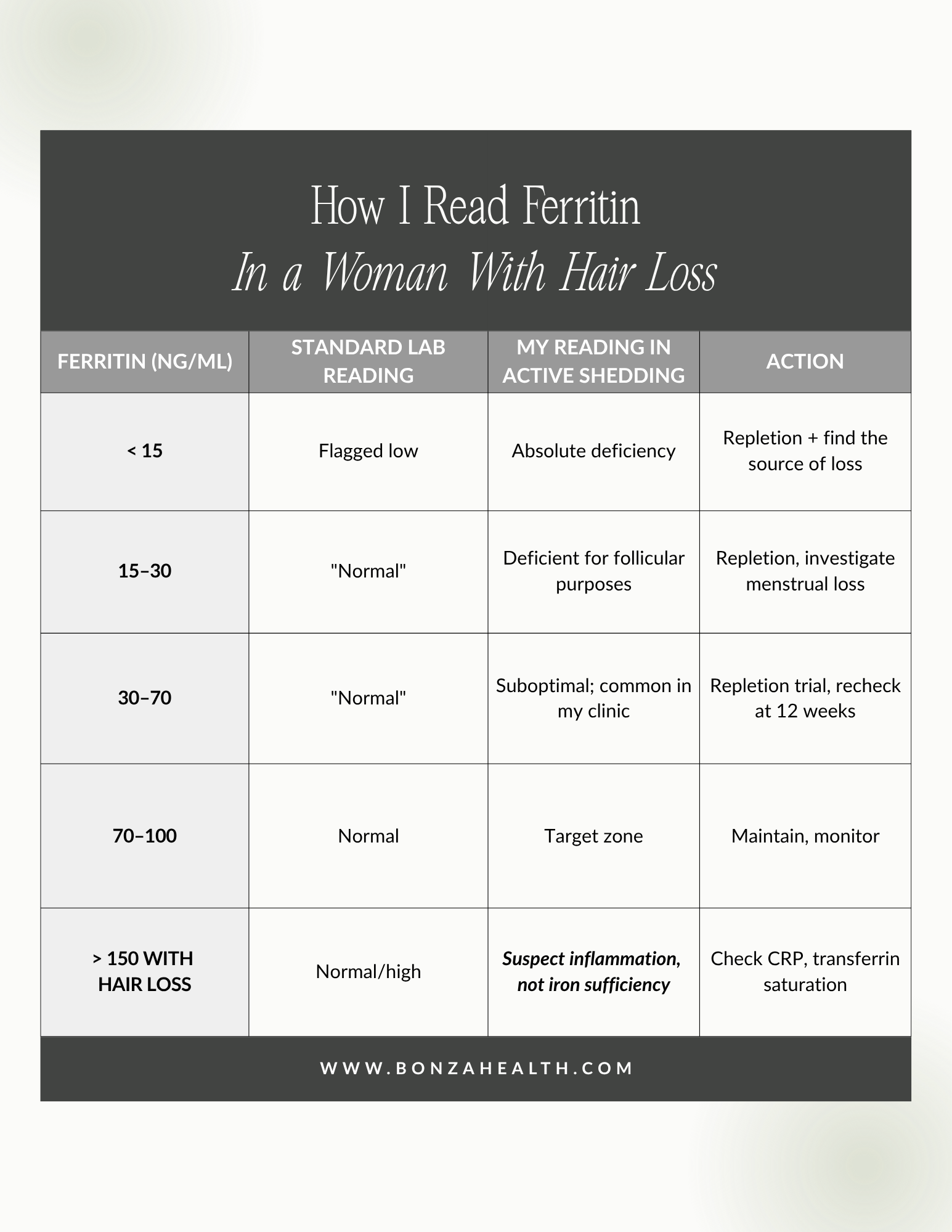
Table 1. How I read ferritin in a woman with hair loss
The confound you must not miss: ferritin is an acute-phase reactant. Infection, obesity, autoimmune disease, alcohol use, and hepatic inflammation all raise it. A woman with a ferritin of 90 and a CRP of 12 may be genuinely iron deficient with a falsely reassuring number. This is why I never order ferritin alone. I order ferritin, CRP, serum iron, TIBC, and transferrin saturation together. A transferrin saturation below 20% with a "normal" ferritin is a red flag that the ferritin is lying.
Oral iron vs. infusion: who qualifies, and why pills fail
Oral iron fails constantly, and it is rarely the patient's fault.
Hepcidin. A single oral iron dose raises hepcidin, the hormone that shuts down intestinal iron absorption, for roughly 24 hours. This means daily — and especially twice-daily — dosing actively suppresses absorption of the next dose. Alternate-day, single-dose regimens produce higher fractional absorption than consecutive daily dosing [7,8]. Most women were told to take iron twice a day with breakfast and dinner. That schedule is close to physiologically self-defeating.
Tolerance. Constipation, nausea, and epigastric pain drive discontinuation before the twelve to twenty-four weeks required to refill stores.
Arithmetic. Maximum daily iron absorption in a depleted woman is roughly 3–5 mg. A woman in perimenopause with anovulatory cycles and heavy, unpredictable bleeding can lose 40 mg or more of iron per cycle. If ongoing loss exceeds maximum absorption, oral repletion cannot mathematically succeed — you are bailing a boat with a hole in it.
That last point is specifically a midlife problem. As ovulation becomes erratic in perimenopause, estrogen goes unopposed by progesterone, the endometrium thickens, and bleeding becomes heavier and less predictable. Many women normalize this. It is the single most common reversible driver of iron depletion I see in women aged 38 to 50.
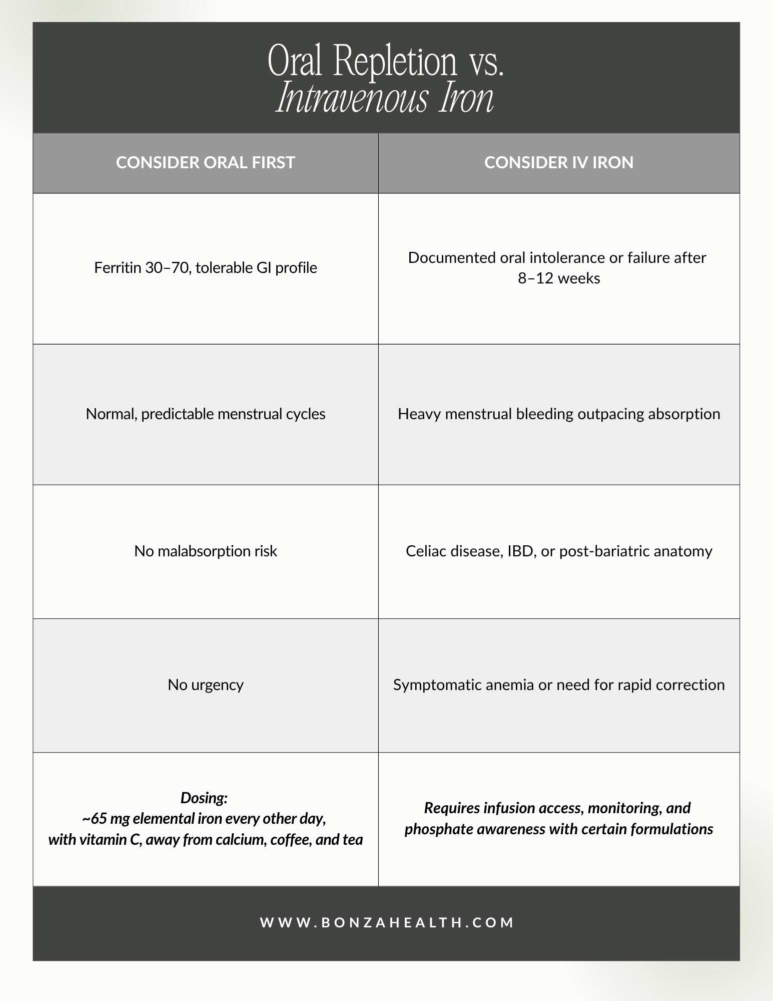
Table 2. Oral repletion vs. intravenous iron
Recheck ferritin at 12 weeks. Visible hair improvement lags biochemical correction by three to six months, because that is how long the follicle cycle takes. I tell every patient this in advance so that month two does not feel like failure.
Copper: the mineral nobody checked
Copper is the quiet partner in the iron story, and aggressive zinc supplementation is the most common reason it goes low.
Copper is required for ceruloplasmin and hephaestin, the ferroxidases that convert Fe²⁺ to Fe³⁺ so iron can be loaded onto transferrin and actually delivered. Without adequate copper, you can swallow iron indefinitely and still not mobilize it — a functional iron deficiency that looks like treatment failure. Copper is also a cofactor for lysyl oxidase, essential to the cross-linking that gives the hair shaft its structural integrity. Profound copper deficiency produces pili torti and hypopigmented, fragile hair; the congenital form (Menkes disease) makes the mechanism unmistakable.
Zinc and copper compete for the same intestinal transporters. Sustained zinc intake above the tolerable upper limit of 40 mg/day suppresses copper absorption, and chronic high-dose zinc can produce frank copper deficiency with anemia, neutropenia, and neurologic sequelae [9]. Many women arrive taking 50 mg of zinc daily for "hair, skin, and nails," bought over the counter, for two years.
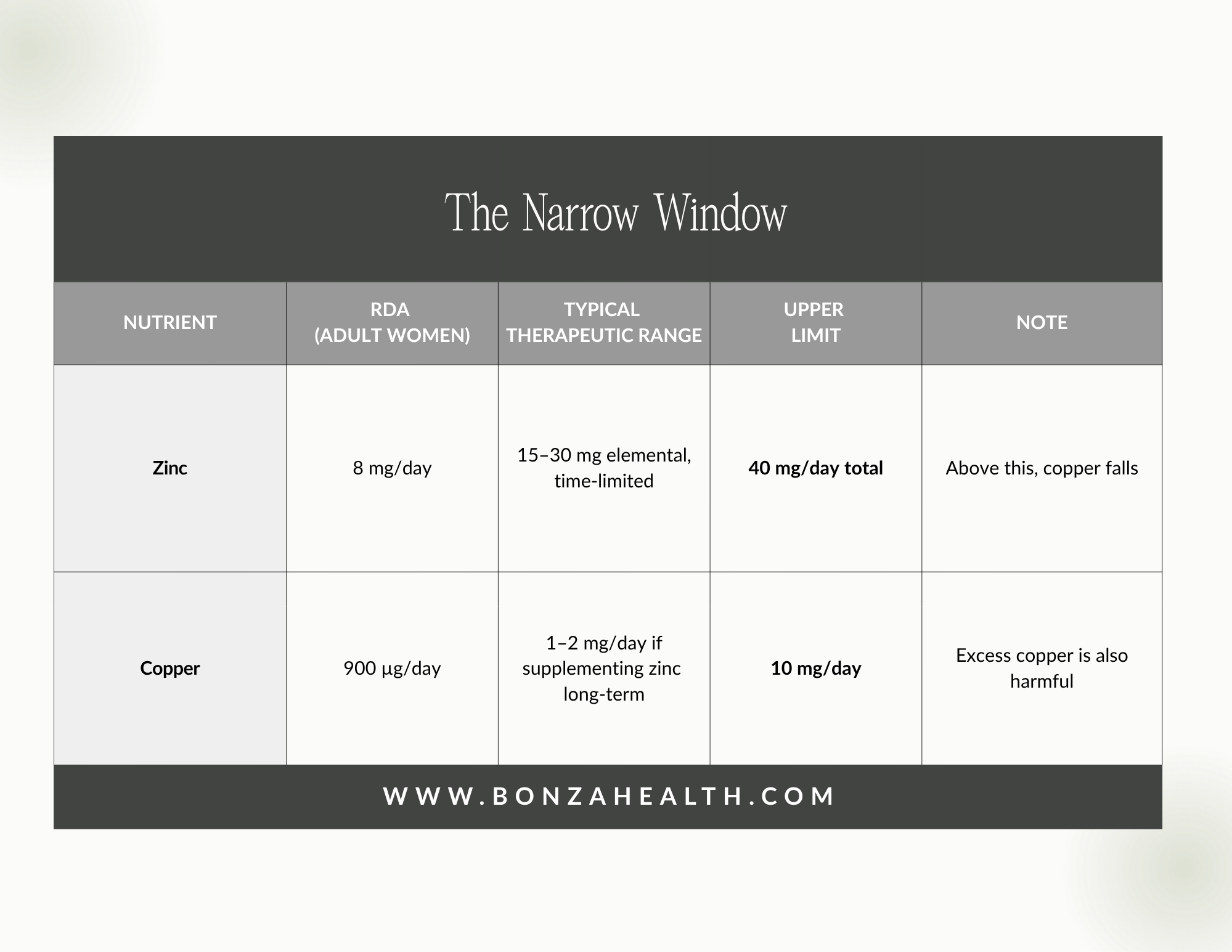
Table 3. The narrow window
The practical rule: if a woman is on therapeutic zinc for more than eight to twelve weeks, copper gets checked and usually gets replaced, and the zinc-to-copper ratio should stay roughly in the 8:1 to 15:1 range.
Thyroid: a normal TSH is not the end of the workup
Thyroid hormone directly governs hair follicle cycling, and hypothyroidism increases the proportion of follicles sitting in telogen. The classic exam finding — loss of the outer third of the eyebrow — is worth knowing, but it is a late sign.
Here is the gap. TSH is a screening test, not a complete thyroid assessment. Autoimmune thyroid disease begins years before TSH leaves the reference range. Thyroid peroxidase antibodies are positive in the overwhelming majority of Hashimoto's thyroiditis, TPOAb prevalence in women of reproductive age runs around 14%, and prevalence rises with age — which places the peak squarely in perimenopause [10]. Antibody-positive women with a TSH in the upper reference range progress to overt hypothyroidism at a substantially higher rate than antibody-negative women.
A woman with a TSH of 3.8, positive TPO antibodies, fatigue, cold intolerance, and diffuse shedding does not have a normal thyroid. She has early autoimmune thyroid disease that has not yet earned a diagnostic code. I test TSH, free T4, free T3, TPOAb, and thyroglobulin antibodies. Whether she needs treatment today is a separate conversation — but she deserves the information, and she deserves monitoring rather than dismissal.
The rest of the panel
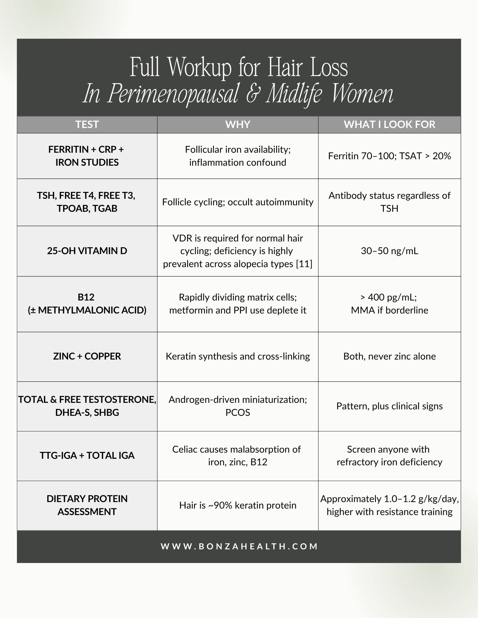
Table 4. Full workup for hair loss in perimenopausal and midlife women
Three history questions carry as much weight as the labs:
Rapid weight loss. Any loss exceeding roughly 10% of body weight over a few months can trigger telogen effluvium two to three months later. This now includes GLP-1 receptor agonists. Pharmacovigilance analysis of the FDA adverse event reporting system found increased reporting odds of alopecia with semaglutide and tirzepatide [12], and a 2025 systematic review confirmed an emerging signal while noting that most reports lack dermatologic confirmation and that causality is unsettled [13]. My clinical read: much of this is telogen effluvium from the rate of loss and the reduced protein and micronutrient intake that accompanies profound appetite suppression — which makes it largely preventable with protein targets and lab monitoring. This is a reason to co-manage, not a reason to stop a medication that is working.
Postpartum timing. Postpartum shedding peaks at three to four months and typically resolves by twelve. Beyond that window, look for depleted iron stores or postpartum thyroiditis.
Crash dieting and restriction. Prolonged low protein intake or severe caloric restriction is a direct cause. If restriction has been driven by distress about weight, that deserves care in its own right.
Visible hair improvement lags biochemical correction by three to six months, because that is how long the follicle cycle takes.
Why a 12-minute appointment cannot do this
Nothing above is exotic. It is not functional medicine or fringe science. It is internal medicine performed at the correct resolution.
But look at what it actually requires: an eight-panel lab draw, a menstrual history detailed enough to estimate blood loss, a supplement inventory including doses, a medication review, a dietary protein assessment, a scalp exam with part-width documentation, and a follow-up structure that can hold twelve weeks of iron repletion and six months of follicle turnover. That is not fifteen minutes. It is a first visit of forty-five to sixty minutes, plus real follow-up.
Most primary care visits are built to move at eleven minutes. In eleven minutes, the honest options are a TSH, a CBC, and reassurance. That is why women in perimenopause keep hearing "your labs are normal" — not because their physicians are careless, but because the system they work inside cannot afford the version of medicine that would find the answer.
That gap is exactly why I practice the way I do.
Frequently Asked Questions
What ferritin level do I need for hair growth?
Most standard labs flag ferritin only below 10–15 ng/mL. For active hair shedding, much of the trichology literature targets above 70 µg/L, with some clinicians aiming toward 100 [1], [3]. This target is clinical judgment supported by observational data, not randomized trial evidence.
Can hair loss be reversed if it's caused by low iron?
Iron-related shedding is a form of telogen effluvium and is generally reversible. Expect three to six months between correcting ferritin and seeing visible density change, because that reflects the hair growth cycle.
Can I have a thyroid problem with a normal TSH?
Yes. Thyroid peroxidase antibodies can be positive for years before TSH becomes abnormal. A complete assessment includes free T4, free T3, and antibodies.
Is perimenopausal hair loss the same as male pattern baldness?
No. Female pattern hair loss typically preserves the frontal hairline and produces diffuse thinning with widening of the central part. It also frequently coexists with a nutritional or thyroid driver — which is the treatable part.
Should I take a hair, skin, and nails supplement?
Not blindly. Many contain zinc at doses that suppress copper over time, and biotin at doses high enough to interfere with troponin and thyroid immunoassays. Test first, supplement second.
Dr. Sarah Bonza is a board-certified physician practicing at Bonza Health. This article is educational and does not constitute individual medical advice. The patient described is a composite drawn from clinical practice, with identifying details changed. To discuss your own workup, visit www.bonzahealth.com.

Sign up for Fullscript to get 10% off professional-grade supplements.
References
[1] D. H. Rushton, "Nutritional factors and hair loss," Clinical and Experimental Dermatology, vol. 27, no. 5, pp. 396–404, Jul. 2002. https://doi.org/10.1046/j.1365-2230.2002.01076.x
[2] J. Kantor, L. J. Kessler, D. G. Brooks, and G. Cotsarelis, "Decreased serum ferritin is associated with alopecia in women," Journal of Investigative Dermatology, vol. 121, no. 5, pp. 985–988, Nov. 2003. https://doi.org/10.1046/j.1523-1747.2003.12540.x
[3] Y. Treister-Goltzman, S. Yarza, and R. Peleg, "Iron deficiency and nonscarring alopecia in women: Systematic review and meta-analysis," Skin Appendage Disorders, vol. 8, no. 2, pp. 83–92, Mar. 2022, https://doi.org/10.1159/000519952
[4] E. A. Olsen, K. B. Reed, P. B. Cacchio, and L. Caudill, "Iron deficiency in female pattern hair loss, chronic telogen effluvium, and control groups," Journal of the American Academy of Dermatology, vol. 63, no. 6, pp. 991–999, Dec. 2010, https://doi.org/10.1016/j.jaad.2009.12.006
[5] D. H. Rushton, W. F. Bergfeld, J. J. H. Gilkes, and D. Van Neste, "Iron deficiency and hair loss—Nothing new?," Journal of the American Academy of Dermatology, vol. 65, no. 1, pp. 203–204, Jul. 2011, https://doi.org/10.1016/j.jaad.2011.02.020
[6] S. A. St. Pierre, G. M. Vercellotti, J. C. Donovan, and M. K. Hordinsky, "Iron deficiency and diffuse nonscarring scalp alopecia in women: More pieces to the puzzle," Journal of the American Academy of Dermatology, vol. 63, no. 6, pp. 1070–1076, Dec. 2010. https://doi.org/10.1016/j.jaad.2009.05.054
[7] D. Moretti et al., "Oral iron supplements increase hepcidin and decrease iron absorption from daily or twice-daily doses in iron-depleted young women," Blood, vol. 126, no. 17, pp. 1981–1989, Oct. 2015. https://doi.org/10.1182/blood-2015-05-642223
[8] N. U. Stoffel et al., "Iron absorption from oral iron supplements given on consecutive versus alternate days and as single morning doses versus twice-daily split dosing in iron-depleted women: Two open-label, randomised controlled trials," The Lancet Haematology, vol. 4, no. 11, pp. e524–e533, Nov. 2017. https://doi.org/10.1016/s2352-3026(17)30182-5
[9] National Institutes of Health, Office of Dietary Supplements, "Zinc: Fact sheet for health professionals," Bethesda, MD, USA. [Tolerable upper intake level, 40 mg/day, adults.] https://ods.od.nih.gov/factsheets/Zinc-HealthProfessional/
[10] American Thyroid Association, "Guidelines for the diagnosis and management of thyroid disease during pregnancy and the postpartum," Thyroid, vol. 27, no. 3, pp. 315–389, 2017. [TPOAb prevalence and progression risk.] https://doi.org/10.1089/thy.2016.0457
[11] "Vitamin D deficiency in non-scarring and scarring alopecias: A systematic review and meta-analysis," Frontiers in Nutrition, 2024. [Reported vitamin D deficiency in 50.4% of female pattern hair loss and 53.5% of telogen effluvium patients.] https://doi.org/10.3389/fnut.2024.1479337
[12] H. Godfrey, Z. Leibovit-Reiben, and P. Jedlowski, "Alopecia associated with the use of semaglutide and tirzepatide: A disproportionality analysis using the FDA adverse event reporting system (FAERS) from 2022 to 2023," Journal of the European Academy of Dermatology and Venereology, vol. 39, no. 2, pp. e153–e154, 2025. https://doi.org/10.1111/jdv.20197
[13] R. F. Rojas Lopez, D. L. Barrera, M. C. Amaya Muñoz, and M. P. Saavedra Diaz, "Alopecia as an emerging adverse effect associated with glucagon-like peptide-1 (GLP-1) receptor agonists for weight loss: A scoping review," Cureus, vol. 17, no. 8, Aug. 2025, https://doi.org/10.7759/cureus.90021