What Every Woman Should Know About Bone Loss, Menopause, and Hormone Therapy
Written and edited by Sarah Bonza, MD, MPH, FAAFP, MSCP, DipABLM, NBC-HWC | Bonza Health
A woman who is still cycling in her late forties or early fifties is frequently in the exact window when bone is lost fastest.
There is a conversation I have had more times than I can count.
A woman in midlife — often still having periods, often somewhere in her forties or fifties — has broken a bone doing something unremarkable. Stepping off a curb. Catching herself on an outstretched hand. Sometimes it has happened more than once. She asks whether her bone density should be checked.
She is told she is too young.
Eventually she gets the scan. The number comes back in the "osteopenia" range — a word that sounds reassuringly minor — and for a while, that number is treated as the answer.
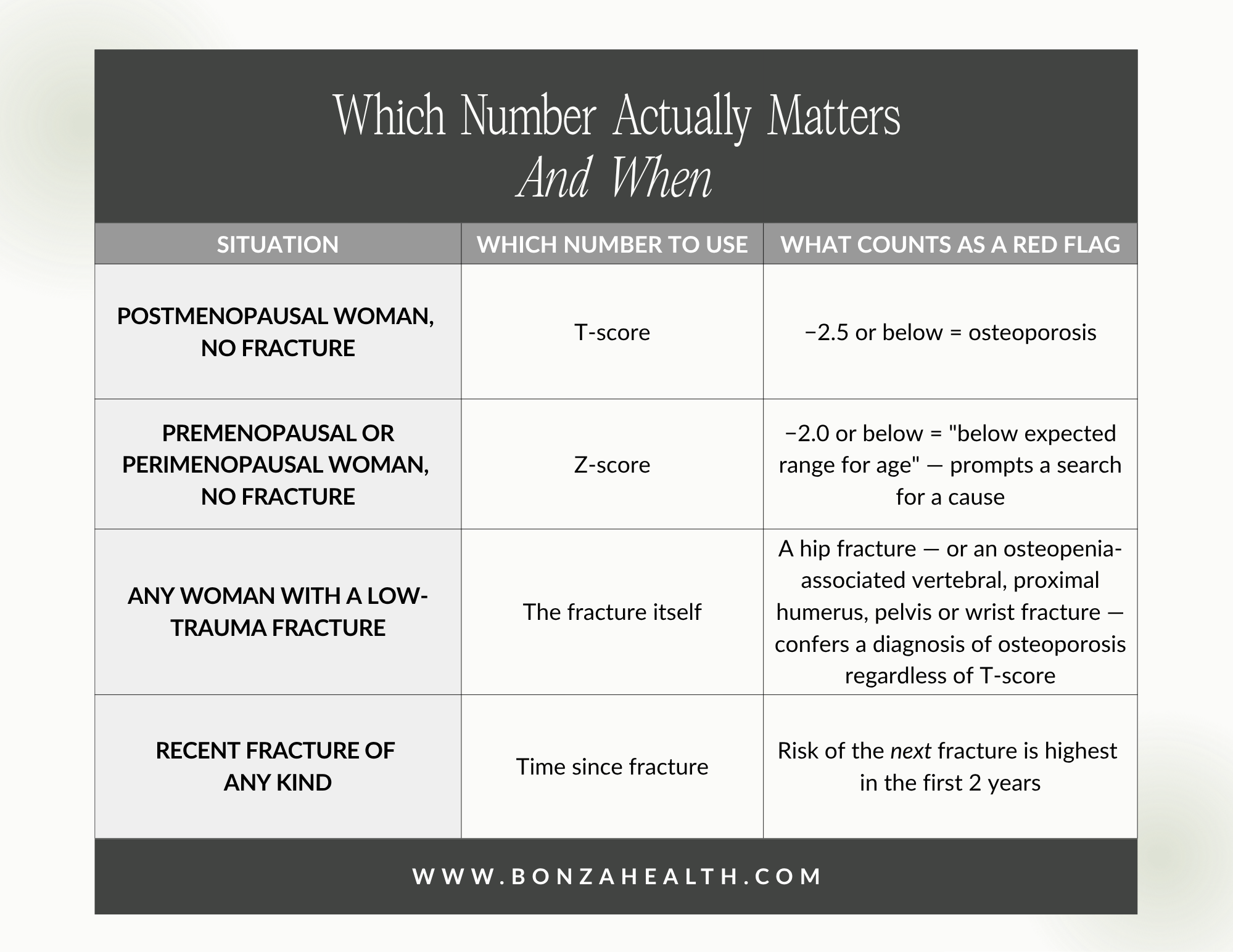
It isn't. When a woman is breaking bones from low-impact injuries, the fractures are the most important information in her chart. Not the T-score.
The scenarios described here reflect patterns I see across many patients. They are not accounts of any individual's medical history.
"Too young for a DEXA" is one of the most expensive sentences in medicine
There is a persistent belief that bone density testing belongs to women in their late sixties and seventies. Screening guidelines reinforce this, and screening guidelines are useful — for screening. They were never designed for a woman who is already breaking.
Once a fragility fracture has happened, we are not screening anymore. We are evaluating an established problem. And the diagnostic rules change accordingly.
The International Working Group on DXA Best Practices makes this explicit: most fragility fractures occur in people whose bone mineral density T-score is above −2.5, and those fractures confirm skeletal fragility even when bone density looks reasonably well preserved [1]. A National Bone Health Alliance working group reached the same conclusion, recommending that a hip fracture, or an osteopenia-associated vertebral, proximal humerus, pelvis, or wrist fracture, should itself confer a diagnosis of osteoporosis [2].
Translation: a normal-ish T-score does not overrule a broken bone. Bone density is one measure of bone strength. It is not the only one, and it is not the best one. The fracture is the outcome we were trying to predict — and it already happened.
There is urgency here, too. Fracture risk is not evenly distributed across the years after a break; it is especially high in the first two years following an incident fracture [3]. Repeated fractures over a short span are not a slow-burning risk profile. They are a fire.
Yet delay remains the norm. In one prospective cohort of women aged 50 and older, 79% had neither been investigated for osteoporosis nor started on anti-fracture therapy six to eight months after a fragility fracture [4]. That is not an individual failing. That is a system-wide pattern — and women who report pain and instability and are told to wait are the ones absorbing the cost.
Sources: [1,2,3]
Perimenopause is not "too early." It's the steepest part of the curve.
The second reason "too young" is so often the wrong answer: a woman who is still cycling in her late forties or early fifties is frequently in the exact window when bone is lost fastest.
Data from the Study of Women's Health Across the Nation (SWAN) mapped this precisely. Bone loss begins about a year before the final menstrual period and decelerates — but does not stop — roughly two years after it [5]. That three-year stretch is now called the transmenopause, and it does the majority of the damage: of a cumulative 10.6% loss at the lumbar spine over ten years, 7.38% occurred during the transmenopause; at the femoral neck, 5.8% of a 9.1% total loss [5]. A separate SWAN analysis found annualized lumbar spine loss beginning roughly two years before the final period at 1.67% per year [6].

Where bone loss actually happens during the menopausal transition

How fast bone disappears during the menopausal transition
So a woman who is still menstruating is not too early for bone assessment. She may be at the single most consequential moment for it.
When bones break young, look for a reason
This is where the picture usually gets more layered — because in younger women, fractures are rarely the whole story.
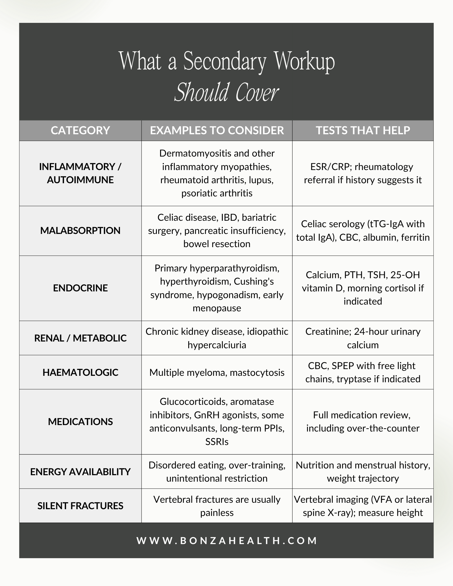
In premenopausal and perimenopausal women, low-trauma fractures are uncommon enough that they demand explanation. The European Calcified Tissue Society and International Osteoporosis Foundation put it plainly: fragility fractures in premenopausal women are rare and mostly secondary — driven by an underlying hormonal, inflammatory, or digestive disorder — and bone density improves when that underlying disease is treated [7]. Broader reviews estimate that secondary causes account for bone loss in more than half of premenopausal and perimenopausal women [8].
Inflammatory and autoimmune disease. Chronic inflammation is directly toxic to bone. In the idiopathic inflammatory myopathies, reported osteoporosis prevalence ranges from 13% to 27%, and vertebral fracture prevalence from 11% to 75% [9]. One cohort found fracture risk and osteoporosis prevalence in myositis comparable to rheumatoid arthritis [10], and a population-based study found raised osteoporosis risk in inflammatory myopathy independent of treatment [11] — meaning the inflammation itself, not only the steroids used to treat it, drives bone loss. Rheumatoid arthritis, lupus, and psoriatic arthritis carry their own skeletal cost. And because autoimmune conditions cluster in families, a first-degree relative with an inflammatory disease is a clinical clue worth following, not a coincidence to note and move past.
Malabsorption. You can eat perfectly and still starve your skeleton. In celiac disease, pooled prevalence among men and premenopausal women is 14.4% for osteoporosis and 39.6% for osteopenia [12], and prospective data show a 30% increase in risk of any fracture and a 69% increase in hip fracture risk [13]. Inflammatory bowel disease carries a pooled odds ratio of 1.32 for osteoporosis [14]. Bariatric surgery, pancreatic insufficiency, and bowel resection belong on the same list.
Endocrine and medication causes. Primary hyperparathyroidism, untreated hyperthyroidism, Cushing's syndrome, hypogonadism, and vitamin D deficiency all merit checking [8]. So do medications: glucocorticoids above all, but also aromatase inhibitors, GnRH agonists, some anticonvulsants, long-term proton pump inhibitors, and SSRIs.
Energy availability. Chronic under-fueling — whether from disordered eating, over-training, or unintentional restriction — suppresses the hormonal signaling bone depends on.
Sources: [7,8,3]
Where hormone therapy fits
My patient came in wanting menopause hormone therapy (MHT), primarily for her bones. That is a legitimate reason to want it, and the evidence supports her instinct.
The Menopause Society's 2022 position statement is unambiguous: hormone therapy remains the most effective treatment for vasomotor symptoms and genitourinary syndrome of menopause, and has been shown to prevent bone loss and fracture. For women under 60 or within 10 years of menopause onset without contraindications, the benefit-risk ratio is favorable for treating bothersome symptoms and preventing bone loss [15].
The underlying data are strong. In the Women's Health Initiative, women randomized to estrogen plus progestin had fewer fractures [16], and conjugated equine estrogen with or without a progestin significantly reduced hip, clinical vertebral, and total fractures [17]. Later WHI analysis found that fracture reduction held regardless of falls risk or baseline FRAX probability [18]. A meta-analysis of 57 randomized trials found a consistent, large, favorable effect on bone density at all sites — a 6.76% difference at the lumbar spine at two years versus control [19].

What hormone therapy does to bone density
Three practical caveats matter enormously:
One: the protection stops when the therapy stops. In WHI, once the intervention was stopped, the fracture benefit dissipated [17]. MHT is not a course of treatment you complete. It is a state you maintain.
Two: route matters. Oral estrogen raises clot risk in a way transdermal estrogen appears not to. A systematic review and meta-analysis found that compared with transdermal estrogen therapy, oral therapy was associated with increased risk of a first venous thromboembolism (RR 1.63), deep venous thrombosis (RR 2.09), and possibly stroke [20]. For a woman whose primary indication is bone, transdermal is often the more sensible starting point.
Three: MHT is not a substitute for finding the cause. If celiac disease or active inflammation is driving the bone loss, estrogen alone treats one variable and leaves the engine running. And for a woman still cycling, MHT requires a regimen that provides adequate endometrial protection — and it is not contraception.
The Menopause Society's 2022 position statement is unambiguous: hormone therapy remains the most effective treatment for vasomotor symptoms and genitourinary syndrome of menopause, and has been shown to prevent bone loss and fracture.
What the Menopause Society recommends
The 2021 osteoporosis position statement lays out a clear framework [3].
The foundation applies to everyone: adequate protein, calcium, and vitamin D; regular physical activity; not smoking; limiting alcohol. Target roughly 1,000–1,200 mg of calcium and 400–800 IU of vitamin D daily, preferentially from diet, with supplements filling gaps rather than replacing food [3]. Exercise is not a soft recommendation: the LIFTMOR trial showed that supervised high-intensity resistance and impact training improved bone density and physical function in postmenopausal women with osteopenia and osteoporosis, with high compliance and only one minor adverse event [21].
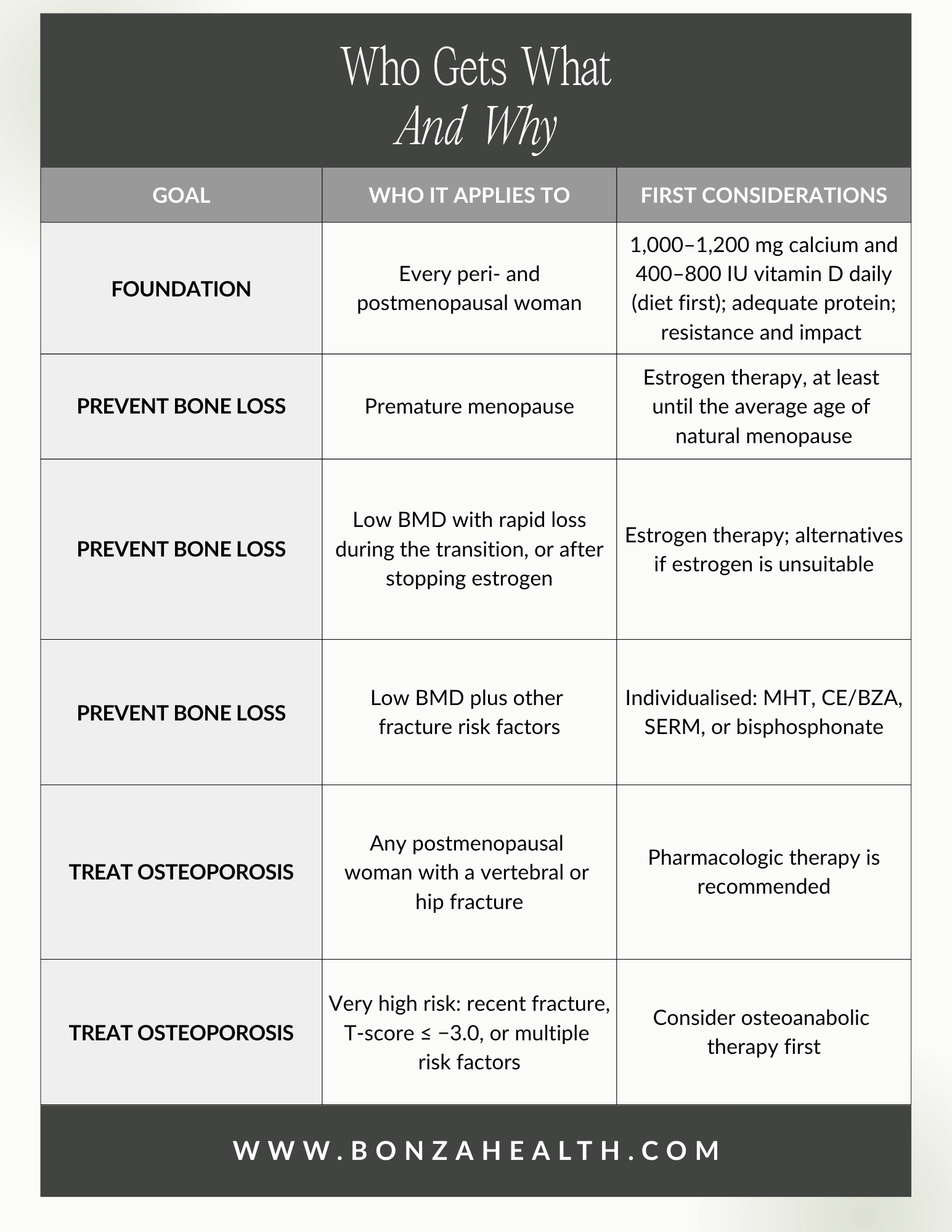
Source: [3]
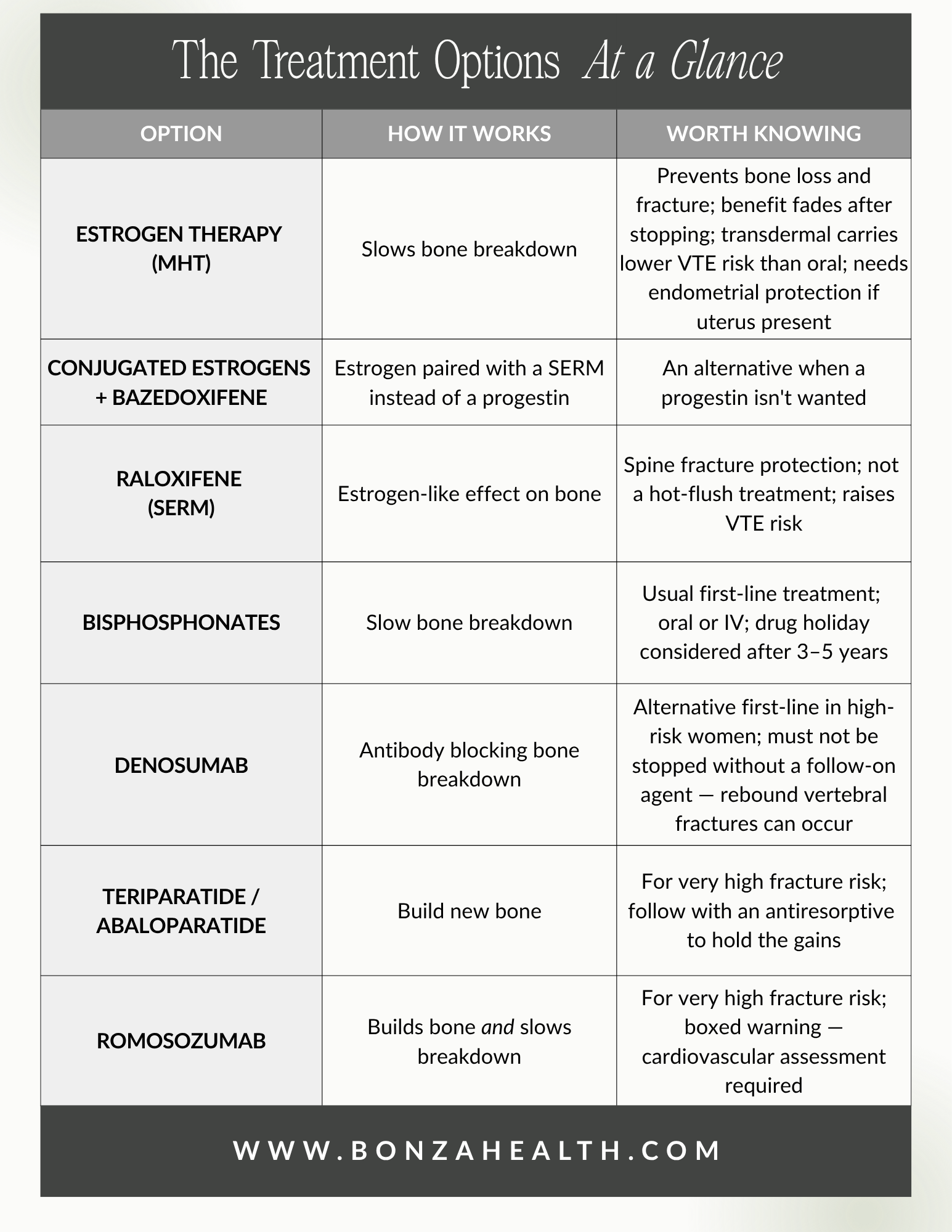
Sources: [3, 22, 23]
When a woman presents with recent fragility fractures, a possible inflammatory contribution, and high near-term risk, the honest answer is usually that hormone therapy alone will not be enough. The plan in that situation typically combines several things at once: a full secondary workup, bone-targeted therapy matched to her fracture burden, hormone therapy where appropriate for symptoms and skeletal support, protein and vitamin D optimization, and supervised loading. Not either/or. Both.
What to ask for
If you have broken a bone doing something ordinary, or lost height, or have a family history of osteoporosis, autoimmune disease, or celiac disease, these are reasonable requests:
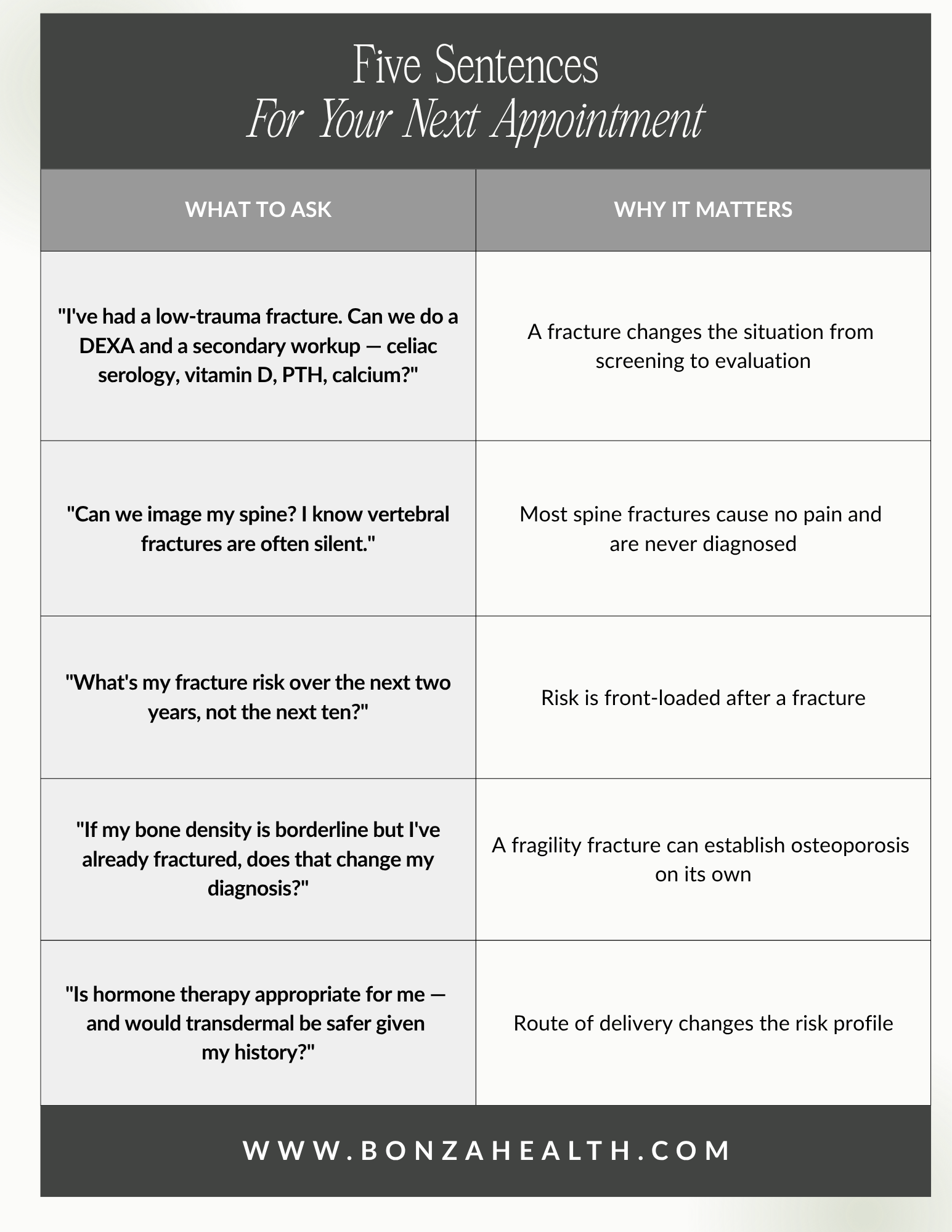
Five sentences for your next appointment
And if you are told you are too young — ask what age your bones think you are.
This article is educational and does not replace individualized medical care. Discuss your own situation with your clinician.

Book your consultation at Bonza Health to learn more about protecting your bone health during perimenopause.
References
[1] A. A. Khan, R. H. J. A. Slart, D. S. Ali, et al., "Osteoporotic fractures: diagnosis, evaluation, and significance from the International Working Group on DXA Best Practices," Mayo Clin. Proc., vol. 99, no. 7, pp. 1127–1141, Jul. 2024. https://doi.org/10.1016/j.mayocp.2024.01.011. PMID: 38960497.
[2] E. S. Siris, R. Adler, J. Bilezikian, et al., "The clinical diagnosis of osteoporosis: a position statement from the National Bone Health Alliance Working Group," Osteoporos. Int., vol. 25, no. 5, pp. 1439–1443, May 2014. https://doi.org/10.1007/s00198-014-2655-z.
[3] The North American Menopause Society, "Management of osteoporosis in postmenopausal women: the 2021 position statement of The North American Menopause Society," Menopause, vol. 28, no. 9, pp. 973–997, Sep. 2021. https://doi.org/10.1097/gme.0000000000001831. PMID: 34448749.
[4] "The care gap in diagnosis and treatment of women with a fragility fracture," Osteoporos. Int., vol. 19, no. 1, pp. 79–86, 2008. https://doi.org/10.1007/s00198-007-0426-9. PMID: 17641811.
[5] G. A. Greendale, M. Sowers, W. Han, M.-H. Huang, J. S. Finkelstein, C. J. Crandall, J. S. Lee, and A. S. Karlamangla, "Bone mineral density loss in relation to the final menstrual period in a multiethnic cohort: results from the Study of Women's Health Across the Nation (SWAN)," J. Bone Miner. Res., vol. 27, no. 1, pp. 111–118, Jan. 2012. https://doi.org/10.1002/jbmr.534. PMID: 21976317.
[6] M. R. Sowers, H. Zheng, M. L. Jannausch, D. McConnell, B. Nan, S. Harlow, and J. F. Randolph Jr., "Amount of bone loss in relation to time around the final menstrual period and follicle-stimulating hormone staging of the transmenopause," J. Clin. Endocrinol. Metab., vol. 95, no. 5, pp. 2155–2162, May 2010. https://doi.org/10.1210/jc.2009-0659. PMID: 20215399.
[7] J. Pepe, J.-J. Body, P. Hadji, et al., "Osteoporosis in premenopausal women: a clinical narrative review by the ECTS and the IOF," J. Clin. Endocrinol. Metab., vol. 105, no. 8, Aug. 2020. https://doi.org/10.1210/clinem/dgaa306. PMID: 32453819.
[8] "Secondary osteoporosis: a review of the recent evidence," Endocr. Pract., vol. 12, no. 4, pp. 436–445, 2006. https://doi.org/10.4158/ep.12.4.436. PMID: 16901802.
[9] "Bone health in idiopathic inflammatory myopathies," Autoimmun. Rev., vol. 20, no. 4, 2021. https://doi.org/10.1016/j.autrev.2021.102782. PMID: 33609795.
[10] A. Vincze, L. Bodoki, K. Szabó, M. Nagy-Vincze, O. Szalmás, J. Varga, K. Dankó, J. Gaál, and Z. Griger, "The risk of fracture and prevalence of osteoporosis is elevated in patients with idiopathic inflammatory myopathies: cross-sectional study from a single Hungarian center," BMC Musculoskelet. Disord., vol. 21, no. 1, art. 426, Jul. 2020. https://doi.org/10.1186/s12891-020-03448-2. PMID: 32616032.
[11] C. W. Lee, C. H. Muo, J. A. Liang, et al., "Increased osteoporosis risk in dermatomyositis or polymyositis independent of the treatments: a population-based cohort study with propensity score," Endocrine, vol. 52, no. 1, pp. 86–92, 2016. https://doi.org/10.1007/s12020-015-0756-x. PMID: 26429781.
[12] R. Ganji, M. Moghbeli, R. Sadeghi, G. Bayat, and A. Ganji, "Prevalence of osteoporosis and osteopenia in men and premenopausal women with celiac disease: a systematic review," Nutr. J., vol. 18, no. 1, art. 9, Feb. 2019. https://doi.org/10.1186/s12937-019-0434-6. PMID: 30732599.
[13] K. Heikkilä, J. Pearce, M. Mäki, and K. Kaukinen, "Celiac disease and bone fractures: a systematic review and meta-analysis," J. Clin. Endocrinol. Metab., vol. 100, no. 1, pp. 25–34, Jan. 2015. https://doi.org/10.1210/jc.2014-1858. PMID: 25279497.
[14] D. F. Hidalgo, B. Boonpheng, J. Phemister, J. Hidalgo, and M. Young, "Inflammatory bowel disease and risk of osteoporotic fractures: a meta-analysis," Cureus, vol. 11, no. 9, e5810, Sep. 2019. https://doi.org/10.7759/cureus.5810. PMID: 31720198.
[15] S. S. Faubion, C. J. Crandall, L. Davis, et al., "The 2022 hormone therapy position statement of The North American Menopause Society," Menopause, vol. 29, no. 7, pp. 767–794, Jul. 2022. https://doi.org/10.1097/gme.0000000000002028. PMID: 35797481.
[16] J. A. Cauley, J. Robbins, Z. Chen, et al., "Effects of estrogen plus progestin on risk of fracture and bone mineral density: the Women's Health Initiative randomized trial," JAMA, vol. 290, no. 13, pp. 1729–1738, Oct. 2003. https://doi.org/10.1001/jama.290.13.1729. PMID: 14519707.
[17] "The Women's Health Initiative: hormone therapy and calcium/vitamin D supplementation trials," Curr. Osteoporos. Rep., vol. 11, no. 3, pp. 171–178, 2013. https://doi.org/10.1007/s11914-013-0150-7. PMID: 23842639.
[18] M. Lorentzon, H. Johansson, N. C. Harvey, E. Liu, L. Vandenput, C. J. Crandall, J. A. Cauley, M. S. LeBoff, E. V. McCloskey, and J. A. Kanis, "Menopausal hormone therapy reduces the risk of fracture regardless of falls risk or baseline FRAX probability — results from the Women's Health Initiative hormone therapy trials," Osteoporos. Int., 2022. https://doi.org/10.1007/s00198-022-06483-y. PMID: 35833956.
[19] G. Wells, P. Tugwell, B. Shea, et al., "Meta-analyses of therapies for postmenopausal osteoporosis. V. Meta-analysis of the efficacy of hormone replacement therapy in treating and preventing osteoporosis in postmenopausal women," Endocr. Rev., vol. 23, no. 4, pp. 529–539, Aug. 2002. https://doi.org/10.1210/er.2001-5002. PMID: 12202468.
[20] K. Mohammed, A. M. Abu Dabrh, K. Benkhadra, et al., "Oral vs transdermal estrogen therapy and vascular events: a systematic review and meta-analysis," J. Clin. Endocrinol. Metab., vol. 100, no. 11, pp. 4012–4020, Nov. 2015. https://doi.org/10.1210/jc.2015-2237. PMID: 26544651.
[21] S. L. Watson, B. K. Weeks, L. J. Weis, A. T. Harding, S. A. Horan, and B. R. Beck, "High-intensity resistance and impact training improves bone mineral density and physical function in postmenopausal women with osteopenia and osteoporosis: the LIFTMOR randomized controlled trial," J. Bone Miner. Res., vol. 33, no. 2, pp. 211–220, Feb. 2018. https://doi.org/10.1002/jbmr.3284. PMID: 28975661.
[22] R. Eastell, C. J. Rosen, D. M. Black, A. M. Cheung, M. H. Murad, and D. Shoback, "Pharmacological management of osteoporosis in postmenopausal women: an Endocrine Society clinical practice guideline," J. Clin. Endocrinol. Metab., vol. 104, no. 5, pp. 1595–1622, May 2019. https://doi.org/10.1210/jc.2019-00221. PMID: 30907953.
[23] D. Shoback, C. J. Rosen, D. M. Black, A. M. Cheung, M. H. Murad, and R. Eastell, "Pharmacological management of osteoporosis in postmenopausal women: an Endocrine Society guideline update," J. Clin. Endocrinol. Metab., vol. 105, no. 3, pp. 587–594, Mar. 2020. https://doi.org/10.1210/clinem/dgaa048.